|
Please note: There is an updated form of this web resource with more interactive elements, and new content! Come and visit Pathweb! |
The purpose of this chapter is simply to introduce you to gross pathology and to demonstrate how to describe and appreciate gross morphology i.e. abnormal organs and tissues.
Consider Mdm Rosnah, a 63 year old lady, who presents with a growth in her foot. It has been increasing in size over the last few months, and is starting to interfere with walking. She also feels the foot is becoming rather unsightly.
Q: After taking a detailed history, what would you do next?
A: The next step would be to examine the patient, with the ultimate aim of making an accurate diagnosis. With the history of a growth in the foot, it would be important to think of a possible neoplastic process, or possibly an infectious process causing skin changes, eg. viral wart, fungal infection.
Take a look at the picture. What clues are there that point to the correct diagnosis?
On physical examination, you carefully examine the foot - this is essentially assessing gross pathology!
")
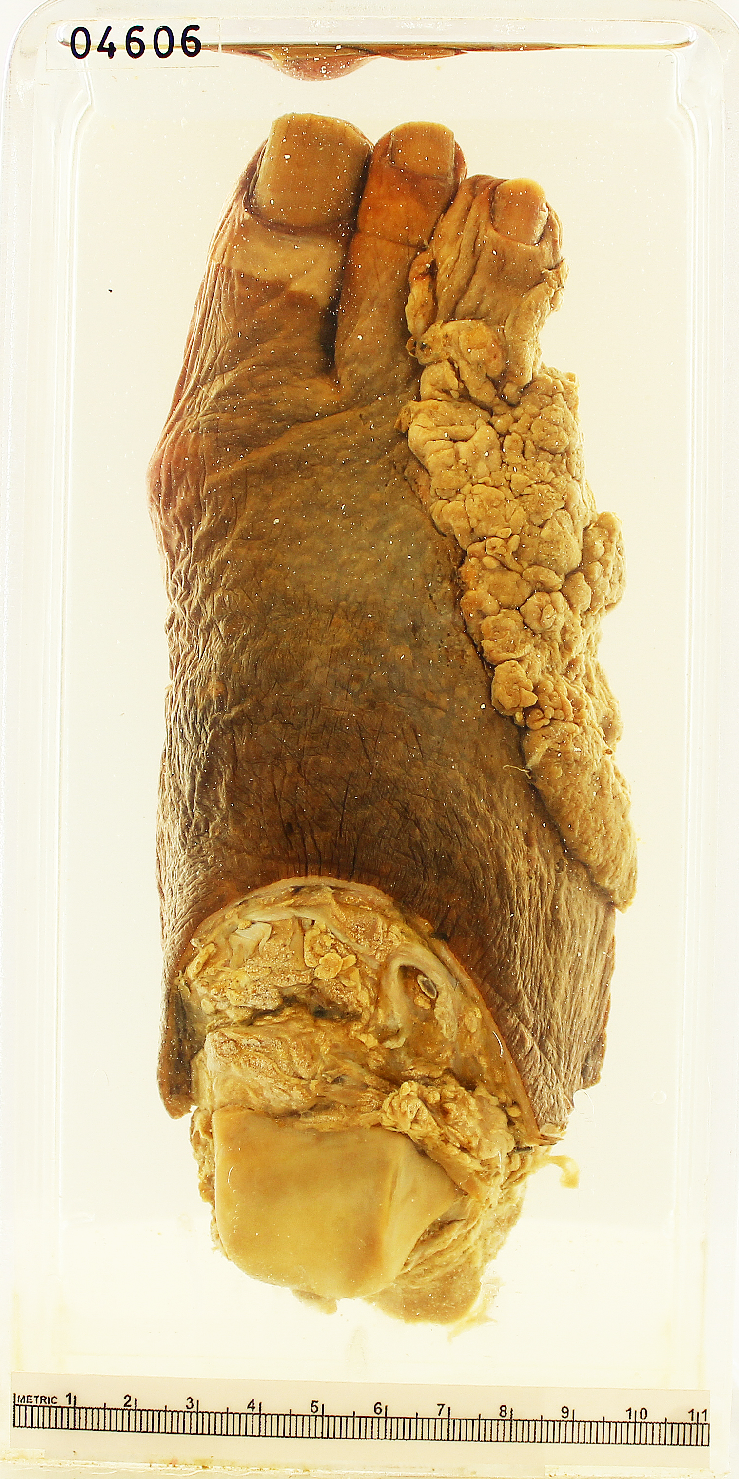
There is a fungating growth arising from the skin of the distal and lateral aspect of the right foot. It has irregular margins and appears to invade into the underlying soft tissue and bone. The last two toes are completely destroyed, while the middle toe shows partial destruction by this growth.
Features are highly suggestive of a malignant tumour.
- Physical examination tells you if this is a tumour, infectious process, or something else. Essentially, the appreciation of the gross pathological features clinches the diagnosis.
- With the correct diagnosis –> one can order appropriate investigations –> proceed to appropriate treatment.
- In internal organs, e.g. in the lung, a radiological test (e.g. X-ray, CT scan) shows the appearance, and this is essentially assessing gross pathology, but in black and white, in vivo.
Key points:
- Essentially, the appreciation of the gross pathological features clinches the diagnosis.
- Subcellular changes –> abnormal appearance of cells –> grossly abnormal organs.
- Gross pathology tells the story of the disease – how it behaves, progresses and gives rise to clinical signs and symptoms.
- In deep seated organs (e.g. liver or lung), we perform radiologic scans or endoscopy – this is the study of gross pathology.
You may find the ONE PAGE Summary useful as a concise summary of the approaches learned. Click on the button below to access it.
.