What is a simple approach to describing gross pathology?
When faced with a pathology specimen, don’t panic –> just be systematic.
There are 5 steps. Here’s what to do: (click on each tab to expand)
Planes:
- Sagittal: Plane that runs down through the body, dividing the body into left and right portions. Subsections of the sagittal plane include:
- Midsagittal runs through the median plane and divides along the line of symmetry
- Parasagittal is parallel to midline but does not divide into equal left and right portions.
- Coronal (frontal): Plane that runs perpendicular to the sagittal plane and divides the body into anterior and posterior (front and back) portions.
- Transverse: Horizontal plane that divides the body into upper and lower portions; also called cross-section.

Transverse section of liver

Coronal section of brain

Sagittal section of knee
- The differential diagnoses (DDx) are quite distinct:
- Diffuse process: the pathology affects the whole organ uniformly
- Enlargement, change in colour, shrinkage/atrophy
- Eg, fatty change in the liver, chronic venous congestion in the lung, diffuse hyperplasia in the thyroid gland
- DDx à Metabolic diseases, autoimmune conditions, haemodynamic disorders, sometimes infections etc.
- Localised/focal process – the pathology is seen as one or several discrete lesions, while the rest of the organ appears grossly normal.
- Mass, cyst, ulcer etc.
- DDx - Neoplasms, cysts, abscesses, infarctions etc.
Nature: mass, nodule, cyst, cavity, scar, focus of discoloration, etc
For mucosal-lined organs eg, Gastrointestinal tract - ulcer, polyp, perforation etc.
“There is a nodule in the upper lobe of the right lung.”
Tip: Try to avoid the word “lesion” – this is entirely non-specific and non-descriptive.
"There is a wedge –shaped, well demarcated area of pallor within the renal cortex in the upper pole of the kidney.”
“The thyroid gland is diffusely enlarged and dark brownish in colour.”
For localized lesions:
- Location – eg. middle lobe of right lung; upper pole of right thyroid lobe, colonic mucosa, subcapsular region of kidney etc.
- Size
- Shape – rounded (eg. benign neoplasms), ovoid, irregular, wedge-shaped (eg. infarcts)
- Number of lesions – solitary, multiple
- Cut surface – texture (solid/cystic/friable), colour (tan, pale, blackish, greenish, variegated), presence of necrosis (paler areas) or haemorrhage
- Edges of lesion
- Solid lesion – well defined vs ill-defined, infiltrative, enc
- Ulcer – regular, punched out, flat edges (favour benign) vs heaped edges, overhanging edges (favour malignant); describe base (necrotic; smooth; visible vessel etc.)
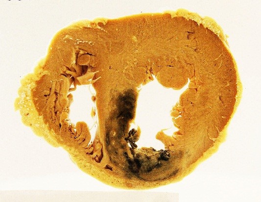
Look for and describe the cause or complication of the disease if shown in the pot – e.g. myocardial infarction with coronary artery thrombus).
In some specimen pots, you may be able to see not only the main pathology but also its cause or complications. Check out the example below.
Exercise: Cause and effect – Below is a heart specimen with a myocardial infarct in the left ventricle. Look carefully = can you spot the cause – the occluded coronary artery (left anterior descending artery)? If not, check out the video below.
We have prepared a glossary of terms for your quick reference. Click on the button below.
.