September 18

Hot Off the Press: Growth differentiation factor-15 and white matter hyperintensities in cognitive impairment and dementia

Vascular pathology plays an important role in the development of cognitive decline and dementia. In this context, growth differentiation factor-15 (GDF-15) has been suggested to be a biomarker due to its regulatory roles in inflammatory and trophic responses during tissue injury. However, limited data exist on the associations of GDF-15 with either cerebrovascular disease (CeVD) burden or the spectrum of cognitive impairment. Therefore, we aimed to study peripheral levels of GDF-15 incognitive impairment no dementia (CIND) or Alzheimer disease (AD) subjects assessed for CeVD using a case–control cohort design, with cases recruited from memory clinics and controls from memory clinics and the community. All subjects underwent detailed neuropsychological assessment, 3-Tesla magnetic resonance imaging, and venous blood draw. Subjects were classified as CIND or AD based on clinical criteria, while significant CeVD was defined as the presence of cortical infarcts and/or 2 lacunes or more, and/or confluent white matter hyperintensities (WMHs) in 2 or more brain regions. A total of 324 subjects were included in the study, of whom 80 had no cognitive impairment, 144 CIND and 100with AD. Higher GDF-15 levels were significantly associated with disease groups, especially in the presence of CeVD, namely, CIND with CeVD (odds ratios [OR]: 7.21; 95% confidence interval [CI]: 2.14–24.27) and AD with CeVD (OR: 21.87; 95% CI: 2.01–237.43). Among the different CeVD markers, only WMH was associated with higher GDF-15 levels (OR: 3.97; 95% CI: 1.79–8.83). The associations between GDF-15 and cognitive impairment as well as with WMH remained significant after excluding subjects with cardiovascular diseases. In conclusion, we showed that increased GDF-15 may be a biomarker for CIND and AD in subjects with WMH.

Reference

Chai YL, Hilal S, Chong JP, Ng YX, Liew OW, Xu X, Ikram MK, Venketasubramanian N, Richards AM, Lai MK, Chen CP (This paper).

February 29

Hot Off the Press: Sphingosine kinase inhibition ameliorates chronic hypoperfusion-induced white matter lesions.

BCAS3

White matter lesions (WML) are thought to contribute to vascular cognitive impairment in elderly patients. Growing evidence show that failure of myelin formation arising from the disruption of oligodendrocyte progenitor cell (OPC) differentiation is a cause of chronic vascular white matter damage. The sphingosine kinase (SphK)/sphingosine-1-phosphate (S1P) signaling pathway regulates oligodendroglia differentiation and function, and is known to be altered in hypoxia. In this study, we measured SphK, S1P as well as markers of WML, hypoxia and OPC (NG2) in a mouse bilateral carotid artery stenosis (BCAS) model of chronic cerebral hypoperfusion. BCAS2
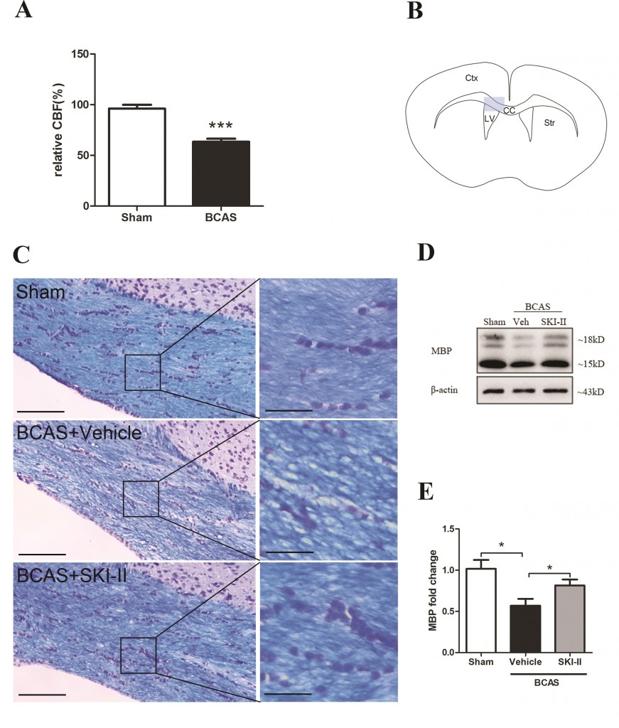
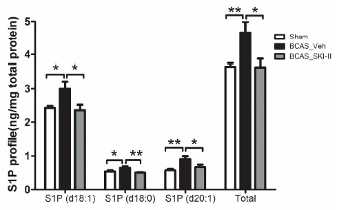
Our results indicated that BCAS induced hypoxia inducible factor (HIF)-1α, Sphk2, S1P, and NG2 up-regulation together with accumulation of WML. In contrast, BCAS mice treated with the SphK inhibitor, SKI-II, showed partial reversal of SphK2, S1P and NG2 elevation and amelioration of WML. In an in vitro model of hypoxia, SKI-II reversed the suppression of OPC differentiation. Our study suggests a mechanism for hypoperfusion-associated WML involving HIF-1α-SphK2-S1P-mediated disruption of OPC differentiation, and proposes the SphK signaling pathway as a potential therapeutic target for white matter disease.

Reference

Yang Y, Torta F, Arai K, Wenk MR, Herr DR, Wong PT, Lai MK (This paper).

February 27

Congratulations, Dr Siew Ying!

Siew Ying 2

Our heartiest congratulations to Siew Ying WONG for successfully defending her thesis. As she heads out and strives on for bigger and better things, we’ll forever be grateful to her for helping establish a rodent model for assessing anti-neuroinflammatory properties of bioactive molecules in our lab. Siew Ying, here’s wishing you every success and fulfillment in the next chapters of your life and work!

February 26

Hot Off the Press: Andrographolide attenuates LPS-stimulated up-regulation of C-C and C-X-C motif chemokines in rodent cortex and primary astrocytes

Andro chemokine brain


Andrographolide is the major bioactive compound isolated from Andrographis paniculata, a native South Asian herb used medicinally for its anti-inflammatory properties. In this study, we aimed to assess andrographolide’s potential utility as an anti-neuroinflammatory therapeutic. Therefore, we conducted an invitro study where the effects of andrographolide on lipopolysaccharide (LPS)-Andro chemokine schematic
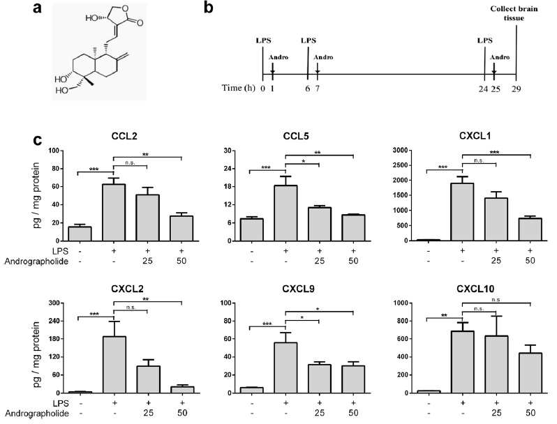
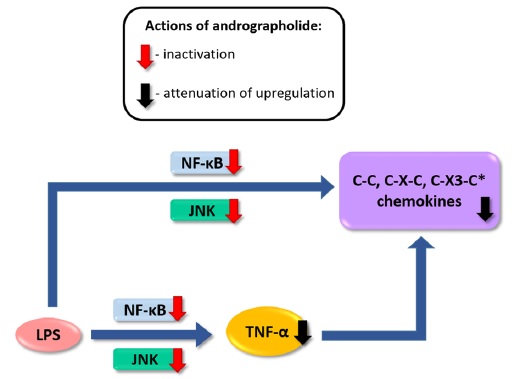
induced chemokine up-regulation both in mouse cortex and in cultured primary astrocytes were measured, including cytokine profiling, gene expression, and, in cultured astrocytes, activation of putative signaling regulators. We found that orally administered andrographolide significantly attenuated mouse cortical chemokine levels from the C-C and C-X-C subfamilies. Similarly, andrographolide abrogated a range of LPS-induced chemokines as well as tumor necrosis factor (TNF)-α in astrocytes. In astrocytes, the inhibitory actions of andrographolide on chemokine and TNF-α up-regulation appeared to be mediated by nuclear factor-κB (NF-κB) or c-Jun N-terminal kinase (JNK) activation. These results suggest that andrographolide may be useful as a therapeutic for neuroinflammatory diseases, especially those characterized by chemokine dysregulation.

Reference

Wong SY, Tan MG, Banks WA, Wong WS, Wong PT, Lai MK (This paper).

February 26

Hot Off the Press: An isoform-specific role of FynT tyrosine kinase in Alzheimer’s disease

Fyn isoform AD

Recent studies suggest that Fyn tyrosine kinase forms part of a toxic triad with β-amyloid and tau in the disease process. However, it is not known whether Fyn is associated with the pathological features of AD in an isoform-specific manner. In this study, we identified selective up-regulation of the alternative-spliced FynT isoform with no change in FynB in the AD neocortex. Furthermore, gene ontology term enrichment analyses and cell type-specific localization of FynT immunoreactivity suggest that FynT up-regulation was associated with neurofibrillary degeneration and reactive astrogliosis. Interestingly, significantly increased FynT in NFT-bearing neurons was concomitant to decreased FynB immunoreactivity, suggesting an involvement of alternative splicing in NFT formation. Furthermore, cultured cells of astrocytic origin have higher FynT to FynB ratio compared to those of neuronal origin. Lastly, primary rat mixed neuron-astrocyte cultures treated with Aβ25-35 showed selective up-regulation of FynT expression in activated astrocytes. Our findings point to an isoform-specific role of FynT in modulating neurofibrillary degeneration and reactive astrogliosis in AD. Fyn kinase is known to interact with β-amyloid and tau, and contributes to Alzheimer’s disease pathogenesis. In this study, it is shown that the alternatively spliced FynT isoform is specifically up-regulated in the AD neocortex, with no change in FynB isoform. The increased FynT correlated with markers of neurofibrillary degeneration and reactive astrogliosis. In primary mixed cultures, treatment with amyloid peptides specifically up-regulated FynT in activated astrocytes. This study points to altered alternative splicing as a potential pathogenic mechanism in AD.

Fyn pTau

Reference

Lee C, Low CY, Francis PT, Attems J, Wong PT, Lai MK, Tan MG (This paper).

February 25

Hot Off the Press: Altered relaxin family receptors RXFP1 and RXFP3 in the neocortex of depressed Alzheimer’s disease patients

RXFP AD

The G-protein-coupled relaxin family receptors RXFP1 and RXFP3 are widely expressed in the cortex and are involved in stress responses and memory and emotional processing. However, the identification of these receptors in human cortex and their status in Alzheimer’s disease (AD), which is characterized by both cognitive impairments and neuropsychiatric behaviours, have not been reported. In this study, we characterized RXFP receptors for immunoblotting and measured RXFP1 and RXFP3 immunoreactivities in the postmortem neocortex of AD patients longitudinally assessed for depressive symptoms. RXFP1 and RXFP3 antibodies were characterized by immunoblotting with lysates from transfected HEK cells and preadsorption with RXFP3 peptides. Also, postmortem neocortical tissues from behaviourally assessed AD and age-matched controls were processed for immunoblotting with RXFP1 and RXFP3 antibodies. Compared to controls, putative RXFP1 immunoreactivity was reduced in parietal cortex of non-depressed AD patients but unchanged in depressed patients. Furthermore, RXFP3 immunoreactivity was increased only in depressed AD patients. RXFP1 levels in the parietal cortex also correlated with severity of depression symptoms. In contrast, RXFP1 and RXFP3 levels did not correlate with dementia severity or β-amyloid burden. Our study suggests that alterations of RXFP1 and RXFP3 may be neurochemical markers of depression in AD, and relaxin family receptors warrant further preclinical investigations as possible therapeutic targets for neuropsychiatric symptoms in dementia.

Reference

Lee JH, Koh SQ, Guadagna S, Francis PT, Esiri MM, Chen CP, Wong PT, Dawe GS, Lai MK (This paper).

October 6

Hot Off the Press: Extracellular vesicles are rapidly purified from human plasma by PRotein Organic Solvent PRecipitation (PROSPR)

PROSPR

Extracellular vesicles (EVs) such as exosomes and microvesicles mediate intercellular communication and regulate a diverse range of crucial biological processes. Host cells that are damaged, infected or transformed release biomarker-containing EVs into the peripheral circulation, where they can be readily accessed for use in diagnostic or prognostic testing. However, current methods of EV isolation from blood plasma are complex and often require relatively large sample volumes, hence are inefficient for widespread use in clinical settings. In work led by collaborator Newman Sze, we report a novel and inexpensive method of rapidly isolating EVs from small volumes of human blood plasma by PRotein Organic Solvent PRecipitation (PROSPR). PROSPR encompasses a rapid three-step protocol to remove soluble proteins from plasma via precipitation in cold acetone, leaving the lipid-encapsulated EVs behind in suspension. This generates higher purity EVs that can then be obtained from filtration or classical ultracentrifugation methods. We foresee that PROSPR-based purification of EVs will significantly accelerate the discovery of new disease biomarkers and the characterization of EVs with potential for clinical applications.

Reference

Gallart-Palau X, Serra A, Wong AS, Sandin S, Lai MK, Chen CP, Kon OL, Sze SK (This paper).