January 25

Hot Off the Press: Serum hepatocyte growth factor is associated with small vessel disease in Alzheimer’s dementia

While hepatocyte growth factor (HGF) is known to exert cell growth, migration and morphogenic effects in various organs, recent studies suggest that HGF may also play a role in synaptic maintenance and cerebrovascular integrity. Although increased levels of HGF have been reported in brain and cerebrospinal fluid (CSF) samples of patients with Alzheimer’s disease (AD), it is unclear whether peripheral HGF may be associated with cerebrovascular disease (CeVD) and dementia. In this study, we examined the association of baseline serum HGF with neuroimaging markers of CeVD in a cohort of pre-dementia (cognitive impaired no dementia, CIND) and AD patients. Serum samples from aged, Non-cognitively impaired (NCI) controls, CIND and AD subjects were measured for HGF levels. CeVD (cortical infarcts, microinfarcts, lacunes, white matter hyperintensities (WMH) and microbleeds) were assessed by magnetic resonance imaging (MRI). After controlling for covariates, higher levels of HGF were associated with both CIND and AD. Among the different CeVD MRI markers in CIND and AD, only small vessel disease, but not large vessel disease markers were associated with higher HGF levels. We conclude that serum HGF may be a useful peripheral biomarker for small vessel disease in subjects with cognitive impairment and AD.

Reference

Zhu Y, Hilal S, Chai YL et al. (This paper).

October 18

Hot Off the Press: An iTRAQ-based proteomic analysis reveals dysregulation of neocortical synaptopodin in Lewy body dementias

In collaboration with the proteomics team led by Newman Sze, we reported an iTRAQ-based study on Lewy body dementias, the second most common cause of neurodegenerative dementia in the elderly after Alzheimer’s disease (AD). The two clinical subgroups of Lewy body dementias, namely, dementia with Lewy bodies (DLB) and Parkinson’s disease dementia (PDD), are differentiated by the chronology of cognitive symptoms relative to parkinsonism. At present, there remains a debate on whether DLB and PDD are separate disease entities, or fall within the same spectrum of Lewy body dementias. In this study, we compared the detergent-soluble proteome via an 8-plex isobaric tag for relative and absolute quantitation (iTRAQ) analysis of pooled lysates from the prefrontal cortex (BA9) of DLB (n = 19) and PDD (n = 21) patients matched a priori for amyloid (total Aβ42) burden, semi-quantitative scores for Lewy bodies and neurofibrillary tangles together with age-matched control (n = 21) subjects. A total of 1914 proteins were confidently identified by iTRAQ (false discovery rate = 0%). None of the proteins showed a significant yet opposite regulation in between DLB and PDD when compared to aged controls in the proteomic data set as well as following immunoblot analysis of the pooled and individual lysates involving all 61 subjects. The postsynaptic protein, synaptopodin (SYNPO) was significantly down-regulated in both DLB and PDD subgroups, suggesting a defective synaptic transmission in the demented patients. In conclusion, the largely similar proteome of DLB and PDD matched for amyloid burden suggests that variations in concomitant AD-related pathology, abnormal post-translational modifications or protein-protein interactions, defective intracellular trafficking or misfolding of proteins could play a part in driving the clinically observed differences between these two subgroups of Lewy body dementias. This further indicates that amyloid-targeting therapeutic strategies may show different efficacies in DLB versus PDD.

Reference

Datta A, Chai YL, Tan JM, et al. (This paper).

May 27

Hot Off the Press: Serum IL-8 is a marker of white-matter hyperintensities in patients with Alzheimer’s disease

First authored by post-graduate student Julie Zhu, we report here that Serum IL-8 may have clinical utility as a biomarker for white matter hyperintensities (WMH) in AD. This is the latest work in our ongoing efforts to examine serum inflammatory markers in preclinical stages of dementia and in AD, as well as to investigate their associations with concomitant cerebrovascular disease (CeVD), given that CeVD have been implicated in cognitive impairment and may worsen AD severity (Zekry et al., 2002). We performed a cross-sectional case-control study including 96 AD, 140 cognitively impaired no dementia (CIND), and 79 noncognitively impaired participants. All subjects underwent neuropsychological and neuroimaging assessments, as well as collection of blood samples for measurements of serum samples interleukin (IL)-6, IL-8, and tumor necrosis factor α levels. Subjects were classified as CIND or dementia based on clinical criteria. Significant CeVD, including white-matter hyperintensities (WMHs), lacunes, and cortical infarcts, was assessed by magnetic resonance imaging. After controlling for covariates, higher concentrations of IL-8, but not the other measured cytokines, were associated with both CIND and AD only in the presence of significant CeVD (CIND with CeVD: odds ratios [ORs] 4.53; 95% confidence interval [CI] 1.5-13.4 and AD with CeVD: OR 7.26; 95% CI 1.2-43.3). Subsequent multivariate analyses showed that among the types of CeVD assessed, only WMH was associated with higher IL-8 levels in CIND and AD (WMH: OR 2.81; 95% CI 1.4-5.6). Longitudinal follow-up studies would help validate these findings.

References

Zhu Y, Chai YL, Hilal S, et al. (This paper).

Zekry D, Duyckaerts C, Moulias R, et al. Degenerative and vascular lesions of the brain have synergistic effects in dementia of the elderly. Acta Neuropathol 103:481-7.

September 18

Hot Off the Press: Growth differentiation factor-15 and white matter hyperintensities in cognitive impairment and dementia

Vascular pathology plays an important role in the development of cognitive decline and dementia. In this context, growth differentiation factor-15 (GDF-15) has been suggested to be a biomarker due to its regulatory roles in inflammatory and trophic responses during tissue injury. However, limited data exist on the associations of GDF-15 with either cerebrovascular disease (CeVD) burden or the spectrum of cognitive impairment. Therefore, we aimed to study peripheral levels of GDF-15 incognitive impairment no dementia (CIND) or Alzheimer disease (AD) subjects assessed for CeVD using a case–control cohort design, with cases recruited from memory clinics and controls from memory clinics and the community. All subjects underwent detailed neuropsychological assessment, 3-Tesla magnetic resonance imaging, and venous blood draw. Subjects were classified as CIND or AD based on clinical criteria, while significant CeVD was defined as the presence of cortical infarcts and/or 2 lacunes or more, and/or confluent white matter hyperintensities (WMHs) in 2 or more brain regions. A total of 324 subjects were included in the study, of whom 80 had no cognitive impairment, 144 CIND and 100with AD. Higher GDF-15 levels were significantly associated with disease groups, especially in the presence of CeVD, namely, CIND with CeVD (odds ratios [OR]: 7.21; 95% confidence interval [CI]: 2.14–24.27) and AD with CeVD (OR: 21.87; 95% CI: 2.01–237.43). Among the different CeVD markers, only WMH was associated with higher GDF-15 levels (OR: 3.97; 95% CI: 1.79–8.83). The associations between GDF-15 and cognitive impairment as well as with WMH remained significant after excluding subjects with cardiovascular diseases. In conclusion, we showed that increased GDF-15 may be a biomarker for CIND and AD in subjects with WMH.

Reference

Chai YL, Hilal S, Chong JP, Ng YX, Liew OW, Xu X, Ikram MK, Venketasubramanian N, Richards AM, Lai MK, Chen CP (This paper).

October 6

Hot Off the Press: Extracellular vesicles are rapidly purified from human plasma by PRotein Organic Solvent PRecipitation (PROSPR)

PROSPR

Extracellular vesicles (EVs) such as exosomes and microvesicles mediate intercellular communication and regulate a diverse range of crucial biological processes. Host cells that are damaged, infected or transformed release biomarker-containing EVs into the peripheral circulation, where they can be readily accessed for use in diagnostic or prognostic testing. However, current methods of EV isolation from blood plasma are complex and often require relatively large sample volumes, hence are inefficient for widespread use in clinical settings. In work led by collaborator Newman Sze, we report a novel and inexpensive method of rapidly isolating EVs from small volumes of human blood plasma by PRotein Organic Solvent PRecipitation (PROSPR). PROSPR encompasses a rapid three-step protocol to remove soluble proteins from plasma via precipitation in cold acetone, leaving the lipid-encapsulated EVs behind in suspension. This generates higher purity EVs that can then be obtained from filtration or classical ultracentrifugation methods. We foresee that PROSPR-based purification of EVs will significantly accelerate the discovery of new disease biomarkers and the characterization of EVs with potential for clinical applications.

Reference

Gallart-Palau X, Serra A, Wong AS, Sandin S, Lai MK, Chen CP, Kon OL, Sze SK (This paper).

August 22

Hot Off the Press: Autoantibodies to GM1 and GQ1bα are not Biological Markers of Alzheimer’s Disease

Uncovering novel biomarkers which lead to better diagnosis, prognosis and treatment is undoubtedly an important endeavor, but sometimes, finding what is not a biomarker may be worthwhile as well. Gangliosides are complex ceramide-glycosphingolipids known to play important roles in neuronal development. The GM1 ganglioside binds to β-amyloid and prevents β-sheet conformational changes (Yanagisawa et al., 1995), while GQ1bα and GT1aα are cholinergic neuron-specific gangliosides found to be increased in murine AD models (Ariga et al. 2010; Ariga et al. 2013a). Antibodies to gangliosides have been detected in AD, but the differences compared to controls were too low to be useful for diagnosis (Chapman et al. 1988). Other studies suffered from small patient numbers (n = 4, Hatzinfilippou et al. 2008), or low cut-off values resulting in low specificity (Ariga et al. 2013b).

autoantibodies AD F1

In this study led by Prof. Nobuhiro Yuki, we measured serum levels of anti-ganglioside antibodies in patients with clinically diagnosed AD (n = 22) and vascular dementia (n = 14) and compared them to sera of patients with Guillain-Barré syndrome (GBS) and multifocal motor neuropathy (MMN), diseases where the antibodies are thought to be pathogenic; as well as to normal controls. We found that titers of IgG and IgM anti-GM1, anti-GQ1bα, and anti-GT1aα antibodies did not differ among AD, vascular dementia, and normal controls, and were remarkably lower than those in GBS and MMN. Our results suggest that the anti-ganglioside antibodies are unlikely to be pathogenic in AD or useful as biomarkers.

References

Miura Y, Miyaji K, Chai YL, Chen CL, Lai MK, Yuki N (This paper).

Ariga T, Yanagisawa M, Wakade C, Ando S, Buccafusco JJ, McDonald MP, Yu RK (2010) Ganglioside metabolism in a transgenic mouse model of Alzheimer’s disease: Expression of Chol-1α antigens in the brain. ASN Neuro 2: e00044.

Ariga T, ItokazuY, McDonald MP, Hirabayashi Y, Ando S, Yu RK (2013a) Brain gangliosides of a transgenic mouse model of Alzheimer’s disease with deficiency in GD3-synthase: Expression of elevated levels of a cholinergic-specific ganglioside, GT1aα. ASN Neuro 5: 141-148.

Ariga T, Kubota M, Nakane M, Oguro K, Yu RK, Ando S (2013b) Anti-Chol-1 antigen, GQ1bα, antibodies are associated with Alzheimer’s disease. PLoS One 8: e63326.

Chapman J, Sela BA, Wertman E, Michaelson DM (1988) Antibodies to ganglioside GM1 in patients with Alzheimer’s disease. Neurosci Lett 86: 235-240.

Hatzifilippou E, Koutsouraki E, Banaki T, Traka M, Costa VG, Baloyannis SJ (2008) Antibodies against GM1 in demented patients. Am J Alzheimers Dis Other Demen 23: 274-279.

Yanagisawa K, Odaka A, Suzuki N, Ihara Y (1995) GM1 ganglioside-bound amyloid beta-protein (Aβ): A possible form of preamyloid in Alzheimer’s disease. Nat Med 1: 1062-1066.

May 6

Hot Off the Press: The brain lipidomes of subcortical ischemic vascular dementia and mixed dementia

VaD MIX lipidome

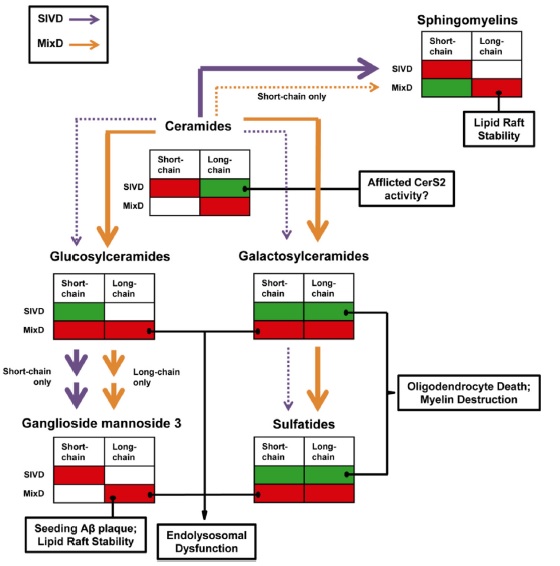
 Despite its importance as the leading cause of vascular dementia in Asia (Chen 2004), the primary pathogenic mechanisms in subcortical ischemic vascular dementia (SIVD) have remained elusive. Because of the lack of approved therapeutic agents for SIVD, there is a pressing need to identify novel therapeutic targets. In work led by collaborators Guanghou Shui and Markus Wenk, we performed comparative lipidomic analyses of SIVD and mixed dementia (i.e., SIVD and Alzheimer’s disease, MixD) which may confer new insights pertaining to the possible interaction between neurodegenerative and vascular mechanisms in the pathogenesis of dementia. Liquid chromatography coupled to mass spectrometry was used to comprehensively analyze the lipidomes of white and gray matter from the temporal cortex of nondemented controls, SIVD, and MixD subjects. Detailed molecular profiles highlighted the pathologic relevance of gray matter sphingolipid fatty acyl chain heterogeneity in dementia. In addition, the levels of sulfatides and lysobisphosphatidic acids were progressively increased in the temporal cortex gray matter from control to SIVD to MixD. White matter phospholipid profiles indicated possible adaptive mechanisms (i.e., increased unsaturation) to chronic ischemia in SIVD and elevated membrane degradation in MixD.

References

Chen CP (2004) Transcultural expression of subcortical vascular disease. J Neurol Sci 226:45-47.

Lam SM, Wang Y, Duan X, Wenk MR, Kalaria RN, Chen CP, Lai MK, Shui G (This paper).

February 11

Hot Off the Press: Novel pathophysiological markers are revealed by iTRAQ-based quantitative clinical proteomics approach in vascular dementia

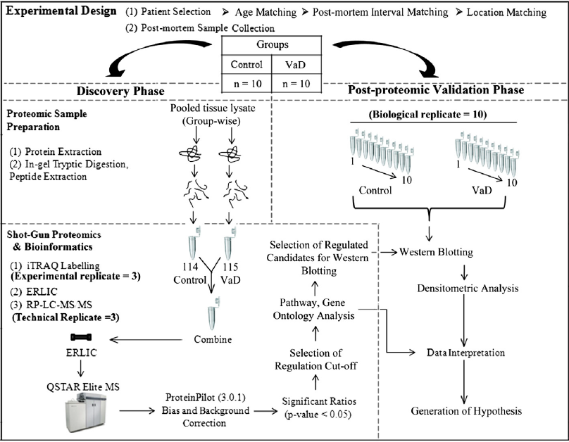
In research led by collaborator Newman Sze, we used an iTRAQ-2D-LC-MS/MS strategy for quantitative analysis of pooled lysates from the neocortex of 21 pathologically confirmed cases of vascular dementia (VaD) and matched non-neurological controls. A total of 144 differentially perturbed proteins out of 2284 confidently identified proteins (false discovery rate=0.3%) were shortlisted for bioinformatics analysis. Western blot analysis of selected proteins using samples from individual patients (n=10 per group) showed significant increases in the abundance of SOD1 and NCAM and reduced ATP5A in VaD. This suggests a state of hypometabolism and vascular insufficiency along with an inflammatory condition during VaD. Elevation of SOD1 and increasing trend for iron-storage proteins (FTL, FTH1) may be indicative of an oxidative imbalance that is accompanied by an aberrant iron metabolism. The synaptic proteins did not exhibit a generalized decrease in abundance (e.g. syntaxin) in the VaD subjects. This reported proteome offers a reference data set for future basic or translational studies on VaD.

VaD Proteomics PNG

Reference

Datta A, Qian J, Chong R, Kalaria RN, Francis P, Lai MK, Chen CP, Sze SK. (This paper).