Imaging and visualization of bone tissue properties

Accurate quantitative estimation of tissue mechanical properties is a research topic. In order for the measurement to be clinical viable, it should be achieved in a non-invasive manner. The estimation of bone density from clinical CT images was reported in 2010 [1]. We are investigating the application of computational intelligent methods on multimodal medical data to aid in the estimation of bone material properties from images. This research has led to the development of opportunistic screening for detection and management of osteopenia [2][3].

There are limited studies on visualization of tissue mechanical properties. However, a visually informative map of the spine (spine invasive map) could provide clinicians with an intuitive representation of the underlying bone properties. Visualization of bone properties can be achieved using material-sensitive transfer functions and coloring schemes that represent different properties:

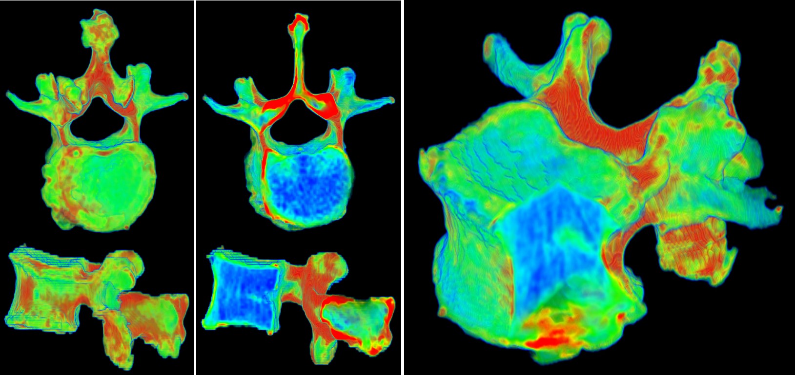
A clustering-based framework for automatic generation of transfer functions for medical visualization was reported in [4].

References:
[1] Zhang, J, CH Yan, CK Chui and SH Ong, “Accurate measurement of bone mineral density using clinical CT imaging with single energy beam spectral intensity correction”. IEEE Transactions on Medical Imaging, 29, 7 (2010): 1382-1389.
[2] Tay, WL, CK Chui, SH Ong and ACM Ng, “Osteopenia screening using areal bone mineral density estimation from diagnostic CT images”. Academic Radiology, 19, no. 10 (2012): 1273-1282.
[3] Tay, WL, CK Chui, SH Ong and ACM Ng, “Ensemble-based regression analysis of multimodal medical data for ostopenia diagnosis”. Expert Systems with Applications, 40, no. 2 (2013): 811-819.
[4] Nguyen, BP, WL Tay, CK Chui and SH Ong, “A Clustering-Based System to Automate Transfer Function Design for Medical Image Visualization”. Visual Computer, 28, no. 2 (2012): 181-191.

Robotic surgery: hand-eye coordination, cognition and biomechanics

Around 2013-2014, I contributed an article with the title above to the  Engineering Research News (ISSN 0217-7870) at the time. The theme was “The Changing Faces of ME”. Mechanical Engineering (ME) is multi-disciplinary.

Following is an edited version:

One key research area pursued by my group is intelligent surgical robotic systems, which augment and enhance the hand-eye coordination capability of the surgeon during operations to achieve the desired outcome and reduce invasiveness.

Hand-eye coordination refers to the ability of our vision system to coordinate and process the information received through the eyes to control, guide and direct our hands in accomplishing a given task. In this work, we studied hand-eye coordination to build a medical simulator for surgical training and to develop medical robot that duplicates the best surgeon’s hand-eye coordination skills.

Our research adopts an integrated view of surgical simulators and robot assisted surgery. The former is a simulation game for surgical training and treatment planning, while the latter involves a single or plurality of devices assisting the surgical team in precise patient operations. With computer simulator, a patient specific surgical plan can be derived with robot manipulation included. By combining patient-specific simulation with robotic execution, we can developed highly autonomous robot(s).

In an automated system, providing proper feedback is crucial to keep the human operator engaged in the decision-making process. Necessary visual, audio and haptic cues should be provided to the human operator in a timely manner, enabling swift intervention. The study on human centricity in an immersive and robot-assisted environment will provide unique insights on human hand-eye coordination capabilities under external influences.

A cognitive engine provides a high level of intelligence in the autonomous robot to be effective collaborator with human(s). The engine possesses knowledge about relevant aspects of surgery, including the dynamics of the surgery, the robot actions and the behavior of biological tissue in response to those actions. The actions of the surgical team contribute to the dynamics and, at times, introduce uncertainty to the operation. The self-learning process of the cognitive engine requires inherent knowledge of tissue biomechanics. Biological tissues within the human patient body cavity are living elements that may be preserved, repaired, or destroyed using mechanical and thermal methods.

Surgery can be planned with a virtual robot in a simulator with realistic biomechanical models, and then the procedure can be performed on the patient using the robot with the assistance of advanced man-machine interfaces.  Augmented reality technologies with intelligent visual, haptic and audio cues will provide a medium for the surgical team to effective control the robot.

The figure depicting the architecture of an intelligent surgical robotic system with a cognitive engine, as mentioned in the original article is still a work-in-progress. Its latest version can be found in:

Tan, X, C B Chng, B Duan, Y Ho, R Wen, X Chen, K B Lim and C K Chui, “Cognitive engine for robot-assisted radio-frequency ablation system”, Acta Polytechnica Hungarica 14, no. 1 (2017): 129-145.

https://uni-obuda.hu/journal/Tan_Chng_Duan_Ho_Wen_Chen_Lim_Chui_72.pdf


Liver tissue properties and frequency-control of RF ablation

A computational model, consisting of an equivalent circuit of resistors and capacitors, is proposed in [1] to investigate the changes in electrical properties of liver tissue during radio-frequency (RF) ablation.  The variations in tissue mechanical properties are correlated with those of the tissue’s electrical properties. RF ablation, in addition to liver tumor treatment, can be utilized to halt blood flow during liver resection. In [2], we further developed the multi-scale model to study the bioimpedance dispersion of liver tissue. The figure below, taken from [3], compares our model with the Cole-Cole model employed in Gabriel’s study. Both models demonstrate a good fit to the experimental data in the high-frequency region. At the lower frequency region, our model provides a better fit to the data.

Using an accurate multi-scale model and a 3D finite element model, we performed RF ablation simulations at various frequencies in [3]. The size of the ablation region increases with higher frequencies. The frequency-control method may prove to be more effective than the duration-control method in RF ablation.

In [4], we previously conducted preliminary work on the application of a multi-scale/multi-level model for simulating molecular medicine through electroporation.

References:

[1] W-H Huang et al. Multi-scale model for investigating the electrical properties and mechanical properties of liver tissue undergoing ablation, Int J CARS (2011) 6:601-607.

[2] W-H Huang et al. A multiscale model for bioimpedance dispersion of liver tissue, IEEE Trans Biomed Eng (2012) 59(6):1593-1597.

[3] B Duan and CK Chui, Multiscale modeling of liver bio-impedance and frequency control for radiofrequency ablation, 2016 IEEE Region 10 Conference (TENCON) – Proceedings of the International Conference, pp. 1532-1535, November 2016.

[4] Chui et al. A medical simulation system with unified multilevel biomechanical model, Proc of 12th International Conference on Biomedical Engineering ICBME 2002, Singapore, 4-7 December 2002.

Constitutive modeling of biological soft tissue

Stress–strain curves of combined porcine liver tissue sample compression and elongation from (Journal of Biomechanics 47 (2014) 2430–2435): (a) Mean values of experimental data, standard deviations from mean values are indicated with horizontal bars; (b) Median values of experimental data; (c) Simulation using the 5-constant Mooney-Rivlin model with parameters calculated by inverse finite element method; and (d) Simulation using the 5-constant Mooney–Rivlin model with parameters calculated by curve fitting.

For modeling and simulating soft tissue indentation, it is important to consider both compression and elongation stress-strain data, as tissue deformation is influenced by both its compressive and tensile characteristics.

An alternative to the Mooney-Rivlin model is a combined logarithmic and polynomial model originally proposed in (Medical & Biological Engineering and Computing 42 (2004) 787-798). The combined logarithmic and polynomial model is superior to the 5-constant Mooney-Rivlin model as the constitutive model for simulation of soft tissue indentation.