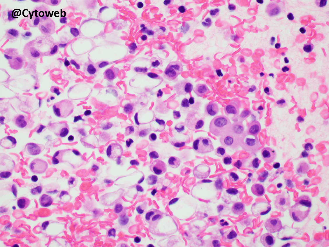
Clinical history: A 72-year-old man with shortness of breath is found to have a pleural effusion. Lung imaging shows consolidation in the ipsilateral lung. A pleural fluid cytology sample is taken, and a cell block processed.

The cells with signet ring morphology are negative for the epithelial marker EP4 and the mesothelial marker WT1. A small group of WT1-positive mesothelial cells is seen. CD163 highlights numerous cells with signet ring cell morphology.
In body cavity fluid samples, the differential diagnoses for cells with signet ring morphology include:
- Metastatic adenocarcinoma with signet ring cell morphology
- Macrophages
- Mesothelial cells
The most helpful morphological features are the nuclei – significant nuclear abnormalities can be seen in adenocarcinoma (hyperchromatic or irregular nuclei with prominent nucleoli) as well as the nature of the cytoplasm (finely bubbly, pinkish in alcohol-stained smears, or containing a small mucin droplet in adenocarcinoma). When the cells contain water as a result of degenerative changes (e.g. in macrophages and mesothelial cells), the vacuole appears clear and empty as seen in this case.
A panel of immunostains will provide the answer as shown above.

The diagnosis is negative: No malignant cells seen. The cells with signet ring morphology are macrophages (CD163-positive, EP4- and WT1-negative). Picture 1 shows the Case of the Month, while picture 2 shows the cell block of a signet ring cell adenocarcinoma. Compare the nuclear and cytoplasmic features.
Case writer: Dr Noel Chia
![]()
GUD CASE THANKS
Good case, very helpful
Thank you