Here are some examples of classical conditions, organised according to categories in The Bethesda System for Reporting Thyroid Cytopathology.
Click on the specific condition to view pic:
Bethesda Category: Classical cases
Benign
Colloid Nodule
Subacute (de Quervain) Thyroiditis
Lymphocytic Thyroiditis
Atypical
Follicular Lesion of Undetermined Significance / Atypia of Undetermined Significance
Follicular Neoplasm
Follicular Neoplasm
Malignant
Papillary Thyroid Carcinoma
Medullary Thyroid Carcinoma
Anaplastic Thyroid Carcinoma
_______________________________________________________
Benign
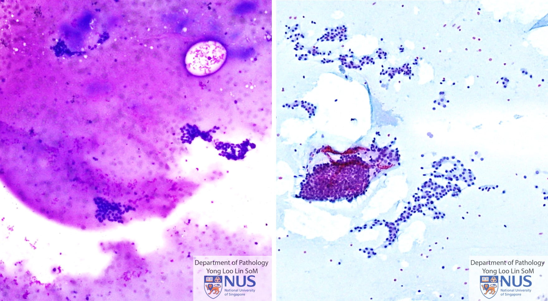


Colloid nodule


- Flat honeycomb sheets of follicular cells which are evenly spaced showing uniform round nuclei with granular chromatin and scant cytoplasm.
- Colloid can appear in the form of a thin, translucent film with bubbles and linear cracks or in the form of thicker blobs.
- Macrophages are sometimes present, and, if abundant or accompanied by granular proteinaceous material, indicate cystic change.
_______________________________________________________


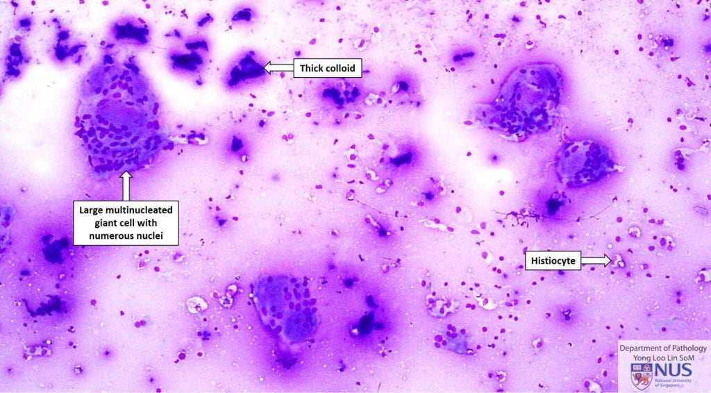


- In addition to follicular cells, there are histiocytes, with varying numbers of lymphocytes and/or some neutrophils.
- May have poorly formed epithelioid granulomas.
- Presence of large multinucleated giant cells containing numerous nuclei is a helpful feature (sometimes these cells engulf colloid).
- Note: Smaller multinucleated giant cells are less specific and may be seen in Hashimoto thyroiditis and papillary thyroid carcinoma.
_______________________________________________________


- Benign groups of follicular cells and scant colloid.
- Follicular cells may show varying proportions of Hurthle cells with round nuclei and abundant granular cytoplasm.
- Many lymphocytes in the background – mixed population, predominantly small lymphocytes.
- Sometimes germinal centre material may be seen. Sometimes multinucleated giant cells are also seen.
_______________________________________________________
Atypical


Atypical; Follicular Lesion of Undetermined Significance / Atypia of Undetermined Significance 


- The FLUS category is a mixed one, covering a spectrum of findings.
- There is architectural and/or nuclear atypia; but not to the extent of being suspicious for malignancy.
- This is an example of FLUS/AUS with architectural atypia:
- In some areas, the follicular cells show architectural atypia e.g. microfollicles, crowded sheets; while in other areas, some flat sheets are seen.
_______________________________________________________
Case 5


Atypical; Follicular Lesion of Undetermined Significance / Atypia of Undetermined Significance 


- The FLUS category is a mixed one, covering a spectrum of findings.
- There is architectural and/or nuclear atypia; but not to the extent of being suspicious for malignancy.
- This is an example of FLUS/AUS with both architectural and nuclear atypia:
- There are crowded sheets of follicular cells arranged in trabecular formations.
- Nuclear atypia is seen, with enlarged nuclei with occasional nuclear grooves and rare nuclear pseudoinclusions.
- There are insufficient definitive diagnostic features for papillary thyroid carcinoma.
_______________________________________________________
Follicular Neoplasm


Follicular Neoplasm 


- Crowded sheets, microfollicles or trabecular arrangements.
- Follicular cells contain round nuclei and with granular chromatin and inconspicuous nucleoli.
_______________________________________________________
Malignant


Malignant; Papillary Thyroid Carcinoma 


- Usually highly cellular aspirates, showing many monolayered syncytial sheets and sometimes papillary structures.
- Nuclei overlap each other within the syncytial sheets.
- Nuclei are often ovoid and enlarged, with fine powdery chromatin, occasional nuclear grooves and nuclear pseudoinclusions.
- Cytoplasm may be delicate or dense.
- Thick colloid (“bubble-gum” colloid), which are better appreciated in Romanowsky stains, may be present.
- Multinucleated giant cells may be noted in the background.
- Sometimes, psammoma bodies may be seen, especially in cystic PTC.
_______________________________________________________


Malignant; Medullary Thyroid Carcinoma 


- Cellular aspirates usually.
- Loose cell clusters and singly occurring cells.
- Cells appear epithelioid/plasmacytoid or spindled.
- Some binucleated or multinucleated cells may be present.
- Nuclei have ‘salt-and-pepper’ chromatin and inconspicuous nucleoli.
- Occasional nuclear inclusions may be seen.
- Sometimes, background amyloid may be identified.
_______________________________________________________


Malignant; Anaplastic Thyroid Carcinoma 


- Markedly pleomorphic nuclei; sometimes spindle cells (sarcomatoid appearance) and squamoid cells.
- Necrosis and inflammatory cells may be seen in the background.